Welcome to the Amira-Avizo Software Use Case Gallery
Below you will find a collection of use cases of our 3D data visualization and analysis software. These use cases include scientific publications, articles, papers, posters, presentations or even videos that show how Amira-Avizo Software is used to address various scientific and industrial research topics.
Use the Domain selector to filter by main application area, and use the Search box to enter keywords related to specific topics you are interested in.
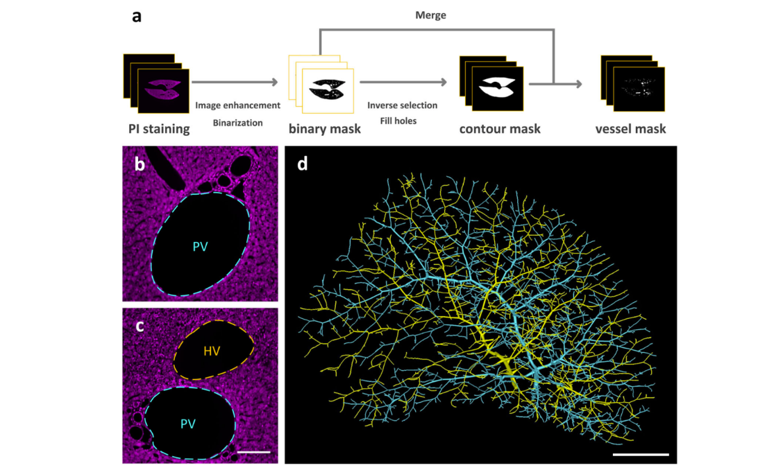
Multiscale reconstruction of various vessels in the intact murine liver lobe
The liver contains a variety of vessels and participates in miscellaneous physiological functions. While past studies generally focused on certain hepatic vessels, we simultaneously obtained all the vessels and cytoarchitectural information of the intact mouse liver lobe at single-cell resolution. […] providing a technology roadmap for studying the fine hepatic vascular structures and their spatial relationship, which will help research into liver diseases and evaluation of medical effi... Read more
Qi Zhang, Anan Li, Siqi Chen, Jing Yuan, Tao Jiang, Xiangning Li, Qingming Luo,Zhao Feng & Hui Gon
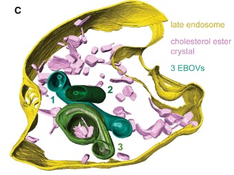
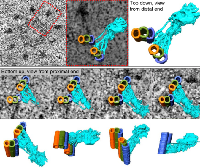
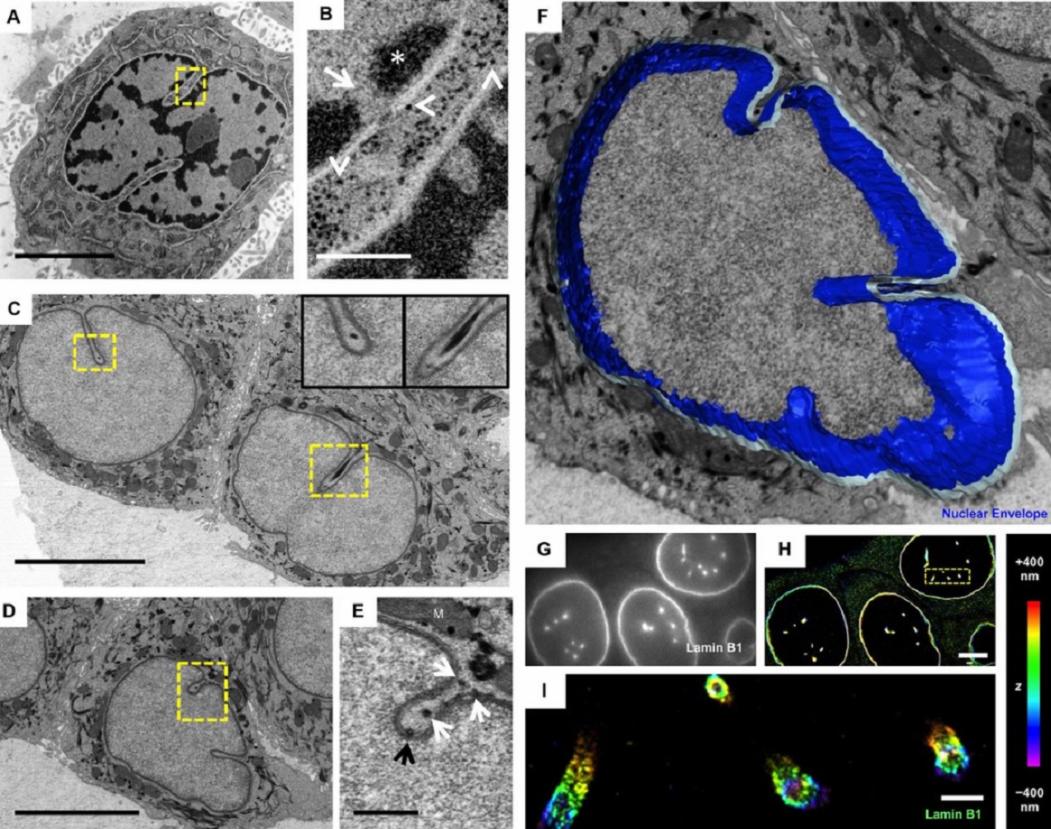
The Ebola virus VP40 matrix layer undergoes endosomal disassembly essential for membrane fusion
Ebola viruses (EBOVs) assemble into filamentous virions, whose shape and stability are determined by the matrix viral protein 40 (VP40). Virus entry into host cells occurs via membrane fusion in late endosomes; however, the mechanism of how the remarkably long virions undergo uncoating, including virion disassembly and nucleocapsid release into the cytosol, remains unknown. Here, we investigate the structural architecture of EBOVs entering host cells and discover that the VP40 matrix disassem... Read more
Sophie L Winter, Gonen Golani, Fabio Lolicato, Melina Vallbracht, Keerthihan Thiyagarajah, Samy Sid Ahmed, Christian Lüchtenborg, Oliver T Fackler, Britta Brügger, Thomas Hoenen, Walter Nickel Ulrich S Schwarz, Petr Chlanda
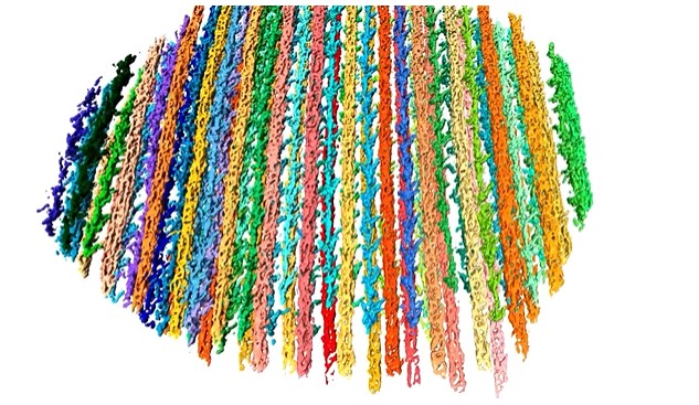
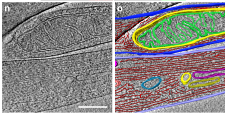
Synergistic role of nucleotides and lipids for the self-assembly of Shs1 septin oligomers
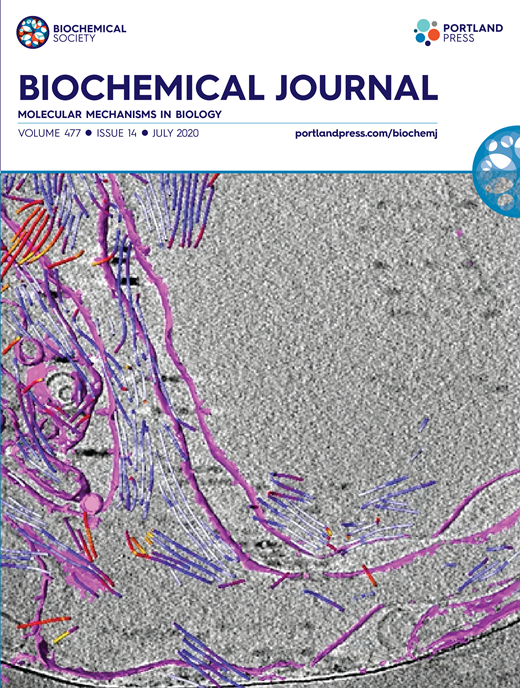
Amira capacities for membranes and filaments segmentation in cryo-TEM images are featured on the front cover of Biochemical Journal, July 2020.
Budding yeast septins are essential for cell division and polarity. (…) [The authors] have dissected, here, for the first time, the behavior of the Shs1 protomer bound to membranes at nanometer resolution, in complex with the other septins. Using electron microscopy, [the authors] have shown that on membranes, Shs1 protomers self-assembl... Read more
Cyntia Taveneau, Rémi Blanc, Gerard Pehau-Arnaudet, Aurélie Cicco, Aurélie Bertin
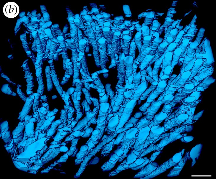
The molecular basis for sarcomere organization in vertebrate skeletal muscle
Sarcomeres are force-generating and load-bearing devices of muscles. A precise molecular picture of how sarcomeres are built underpins understanding their role in health and disease. Here, we determine the molecular architecture of native vertebrate skeletal sarcomeres by electron cryo-tomography.
Our reconstruction reveals molecular details of the three-dimensional organization and interaction of actin and myosin in the A-band, I-band, and Z-disc and demonstrates that α-actinin cros... Read more
Zhexin Wang, Michael Grange, Thorsten Wagner, Ay Lin Kho, Mathias Gautel, Stefan Raunser
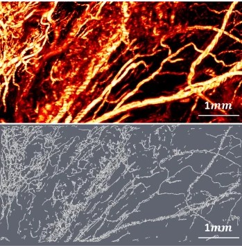
Optical Resolution Photoacoustic Microscopy of Ovary and Fallopian Tube
Ovarian cancer is the leading cause of death among gynecological cancers, but is poorly amenable to preoperative diagnosis. In this study, we investigate the feasibility of “optical biopsy,” using high-optical-resolution photoacoustic microscopy (OR-PAM) to quantify the microvasculature of ovarian and fallopian tube tissue. The technique is demonstrated using excised human ovary and fallopian tube specimens imaged immediately after surgery.
This report describes the first applicatio... Read more
Bin Rao, Xiandong Leng, Yifeng Zeng, Yixiao Lin, Ruimin Chen, Qifa Zhou, Andrea R. Hagemann, Lindsay M. Kuroki, Carolyn K. McCourt, David G. Mutch, Matthew A. Powell, Ian S. Hagemann & Quing Zhu
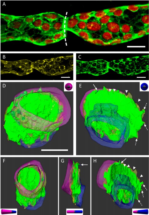
3D Dissection of Structural Membrane-Wall Contacts in Filamentous Moss Protonemata
Cell-to-cell contact is essential for communication and development of multicellular organisms. A prerequisite is the passage through membranes. That way, molecular exchange and information flow is regulated via hormones, membrane proteins and pores.
In plants, the rigid cell walls prevent large membrane contact areas between protoplasts. Only plasmodesmata, minute channels between adjacent cells, form direct connections. Often, molecular data of the proteins involved are manifold but t... Read more
Dominik Harant and Ingeborg Lang
The Architecture of Traveling Actin Waves Revealed by Cryo-Electron Tomography
Actin waves are dynamic supramolecular structures involved in cell migration, cytokinesis, adhesion, and neurogenesis. Although wave-like propagation of actin networks is a widespread phenomenon, the actin architecture underlying wave propagation remained unknown. In situ cryo-electron tomography of Dictyostelium cells unveils the wave architecture and provides evidence for wave progression by de novo actin nucleation. Subtomogram averaging reveals the structu... Read more
Marion Jasnin, Florian Beck, Mary Ecke, Yoshiyuki Fukuda, Antonio Martinez-Sanchez, Wolfgang Baumeister, Günther Gerisch
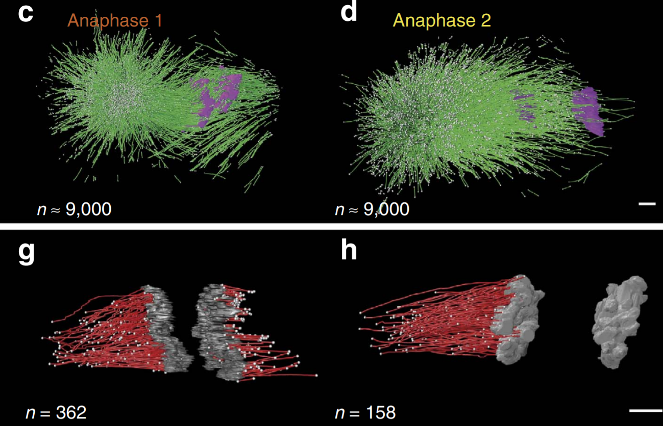
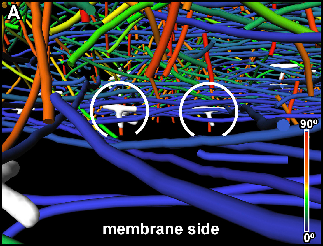
C. elegans chromosomes connect to centrosomes by anchoring into the spindle network
The mitotic spindle ensures the faithful segregation of chromosomes. Here we combine the first large-scale serial electron tomography of whole mitotic spindles in early C. elegans embryos with live-cell imaging to reconstruct all microtubules in 3D and identify their plus- and minus-ends. We classify them as kinetochore (KMTs), spindle (SMTs) or astral microtubules (AMTs) according to their positions, and quantify distinct properties of each class. While our light microscopy and muta... Read more
Stefanie Redemann, Johannes Baumgart, Norbert Lindow, Michael Shelley, Ehssan Nazockdast, Andrea Kratz, Steffen Prohaska, Jan Brugués, Sebastian Fürthauer & Thomas Müller-Reichert
Centrioles are vital cellular structures that form centrosomes and cilia. The formation and function of cilia depends on a set of centriole’s distal appendages. In this study, we use correlative super resolution and electron microscopy to precisely determine where distal appendage proteins localize in relation to the centriole microtubules and appendage electron densities. Here we characterize a novel distal appendage protein ANKRD26 and detail, in high resolution, the initial steps of dist... Read more
Mathew Bowler, Dong Kong, Shufeng Sun, Rashmi Nanjundappa, Lauren Evans, Veronica Farmer, Andrew Holland, Moe R. Mahjoub, Haixin Sui & Jadranka Loncarek

Soluble tubulin is locally enriched at mitotic centrosomes in C. elegans
During mitosis, the centrosome expands its capacity to nucleate microtubules. Understanding the mechanisms of centrosomal microtubule nucleation is, however, constrained by a lack of knowledge of the amount of soluble and polymer tubulin at mitotic centrosomes. Here we combined light microscopy and serial-section electron tomography to measure the amount of dimer and polymer at mitotic centrosomes in early C. elegans embryos. We show that a C. elegans one-cell stage centrosome at metaphase co... Read more
Johannes Baumgart, Marcel Kirchner, Stefanie Redemann, Jeffrey Woodruff, Jean-Marc Verbavatz, Frank Julicher, Anthony Hyman, Thomas Mueller-Reichert, Jan Brugues
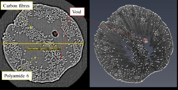
Continuous carbon fibre-reinforced polymer (CCFRP) composites have been increasingly used in airframes and other high-end industrial products due to their superior mechanical properties such as high strength-weight and stiffness-weight ratios […] Additive manufacturing (also known as 3D printing) has shown potential to fabricate continuous fibre-reinforced composites with highly complex shapes in both 2D and 3D. Among them, Fused Filament Fabrication (FFF) 3D printing melts and deposits... Read more
Haoqi Zhang, Jiayun Chen, Dongmin Yang
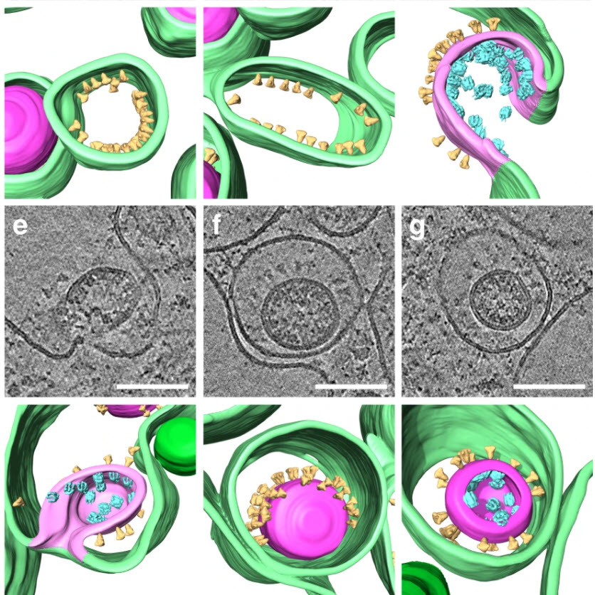
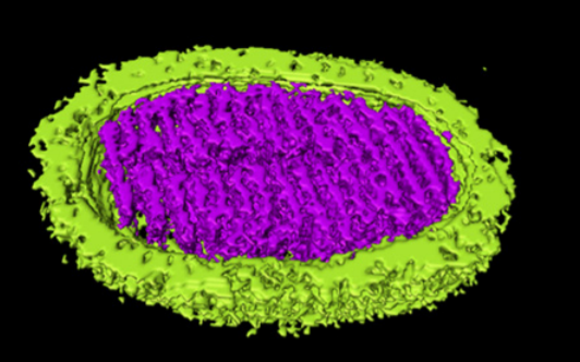
SARS-CoV-2 structure and replication characterized by in situ cryo-electron tomography
Severe acute respiratory syndrome coronavirus 2 (SARS-CoV-2), the causative agent of the COVID19 pandemic, is a highly pathogenic β-coronavirus. As other coronaviruses, SARS-CoV-2 is enveloped, replicates in the cytoplasm and assembles at intracellular membranes. Here, we structurally characterize the viral replication compartment and report critical insights into the budding mechanism of the virus, and the structure of extracellular virions close to their native state by in situ cryo-electr... Read more
Steffen Klein, Mirko Cortese, Sophie L. Winter, Moritz Wachsmuth-Melm, Christopher J. Neufeldt, Berati Cerikan, Megan L. Stanifer, Steeve Boulant, Ralf Bartenschlager, Petr Chlanda
Novel Sulfolobus Virus with an Exceptional Capsid Architecture
A novel archaeal virus, denoted Sulfolobus ellipsoid virus 1 (SEV1), was isolated from an acidic hot spring in Costa Rica. The morphologically unique virion of SEV1 contains a protein capsid with 16 regularly spaced striations and an 11-nm-thick envelope. The capsid exhibits an unusual architecture in which the viral DNA, probably in the form of a nucleoprotein filament, wraps around the longitudinal axis of the virion in... Read more
Haina Wang, Zhenqian Guo, Hongli Feng, Yufei Chen, Xiuqiang Chen, Zhimeng Li, Walter Hernández-Ascencio, Xin Dai, Zhenfeng Zhang, Xiaowei Zheng, Marielos Mora-López, Yu Fu, Chuanlun Zhang, Ping Zhu, Li Huang
The importance of context in regulation of gene expression is now an accepted principle; yet the mechanism by which the microenvironment communicates with the nucleus and chromatin in healthy tissues is poorly understood. A functional role for nuclear and cytoskeletal architecture is suggested by the phenotypic differences observed between epithelial and mesenchymal cells…
Read more
Danielle M. Jorgens, Jamie L. Inman, Michal Wojcik, Claire Robertson, Hildur Palsdottir, Wen-Ting Tsai, Haina Huang, Alexandre Bruni-Cardoso, Claudia S. López, Mina J. Bissell, Ke Xu, Manfred Auer
Correlative cryo-electron microscopy reveals the structure of TNTs in neuronal cells
The orchestration of intercellular communication is essential for multicellular organisms. One mechanism by which cells communicate is through long, actin-rich membranous protrusions called tunneling nanotubes (TNTs), which allow the intercellular transport of various cargoes, between the cytoplasm of distant cells in vitro and in vivo. Here, we use correlative FIB-SEM, light- and cryo-electron microscopy approaches to elucidate the structural organization of neuronal TNTs. Our data indicate ... Read more
Anna Sartori-Rupp, Diégo Cordero Cervantes, Anna Pepe, Karine Gousset, Elise Delage, Simon Corroyer-Dulmont, Christine Schmitt, Jacomina Krijnse-Locker & Chiara Zurzolo
Microbial-tubeworm associations in a 440 million year old hydrothermal vent community
Microorganisms are the chief primary producers within present-day deep-sea hydrothermal vent ecosystems, and play a fundamental role in shaping the ecology of these environments. (…) The oldest known hydrothermal vent community that includes metazoans is preserved within the Ordovician to early Silurian Yaman Kasy massive sulfide deposit, Ural Mountains, Russia. (…) A re-examination of these fossils using a range of microscopy, chemical analysis and nano-tomography techniques re... Read more
Magdalena N. Georgieva , Crispin T. S. Little , Russell J. Bailey , Alexander D. Ball and Adrian G. Glover